Imaging
CT Scanning
The High-Resolution X-ray Computed Tomography Facility (UTCT) is a national shared multi-user facility supported by the Instrumentation and Facilities Program of NSF’s Earth Sciences directorate.
- Geological Applications
- Biological and Paleontological Applications
- Anthropological Applications
- Engineering and Other Applications
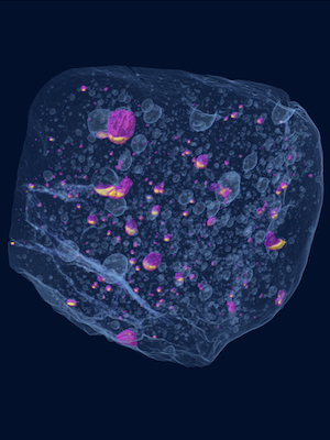
CT allows researchers to visualize and obtain 3D measurements of the interiors of opaque solid objects such as rocks, fossils, and meteorites.
- Contact Richard Ketcham for details.
Optical Microscopy and Mapping
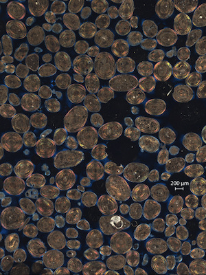
Light microscopes allow micro-scale imaging of rocks, fossils, and other samples with directly observed visible light. Capabilities in the Martindale Research Group include stereo microscopes with image stacking capabilities (depths of focus) with direct and transmitted light (0.5x – 12x magnification), petrographic microscopes with image stacking to capture multiple depths of focus and tiling capabilities allowing users to image entire thin sections (transmitted and reflected light, 1.25x – 500x magnification), and macro-scale photography set ups (angled and polarized light).
- Contact Rowan Martindale for details.
Under Shared Facilities, check out the Graduate and Undergraduate Microscopy Lab
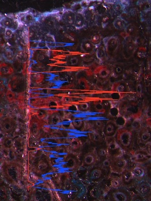
Elemental Mapping
Several labs in the department use ion and electron beam instruments to image samples.
- The laser mode of the ICP-MS in the Quadrapole Lab. Contact Nathan Miller for details.
- The E-beam Lab houses SEM, electron microbeam, and CL. Contact Kenneth Befus for details.
- UT Chron Lab houses a laser ablation ICP-MS as well. Contact Lisa Stockli for details.